CT-CUBE 1 MOBILE CT UNIT

BUILDING SPECIFICATION

BUILDING EQUIPMENT

BUILDING EQUIPMENT

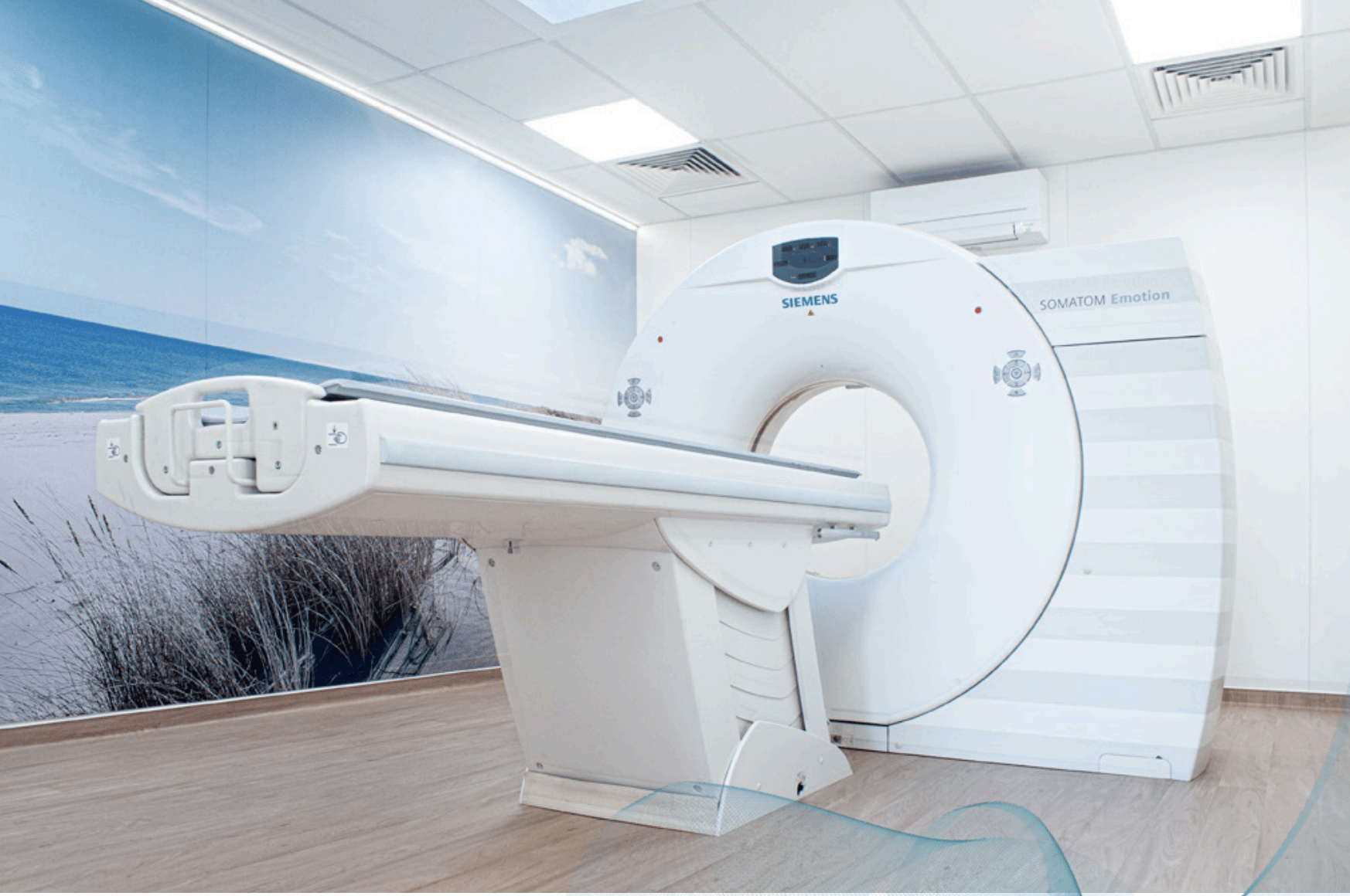
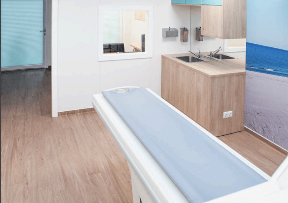
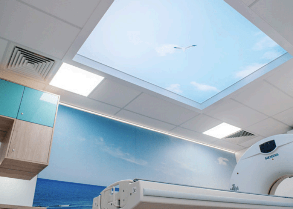
Siemens Somatom Emotion 6 with examination table
Accessories such as positioning aids, phantoms, etc. are easily accessible in the examination room in built-in cupboards.

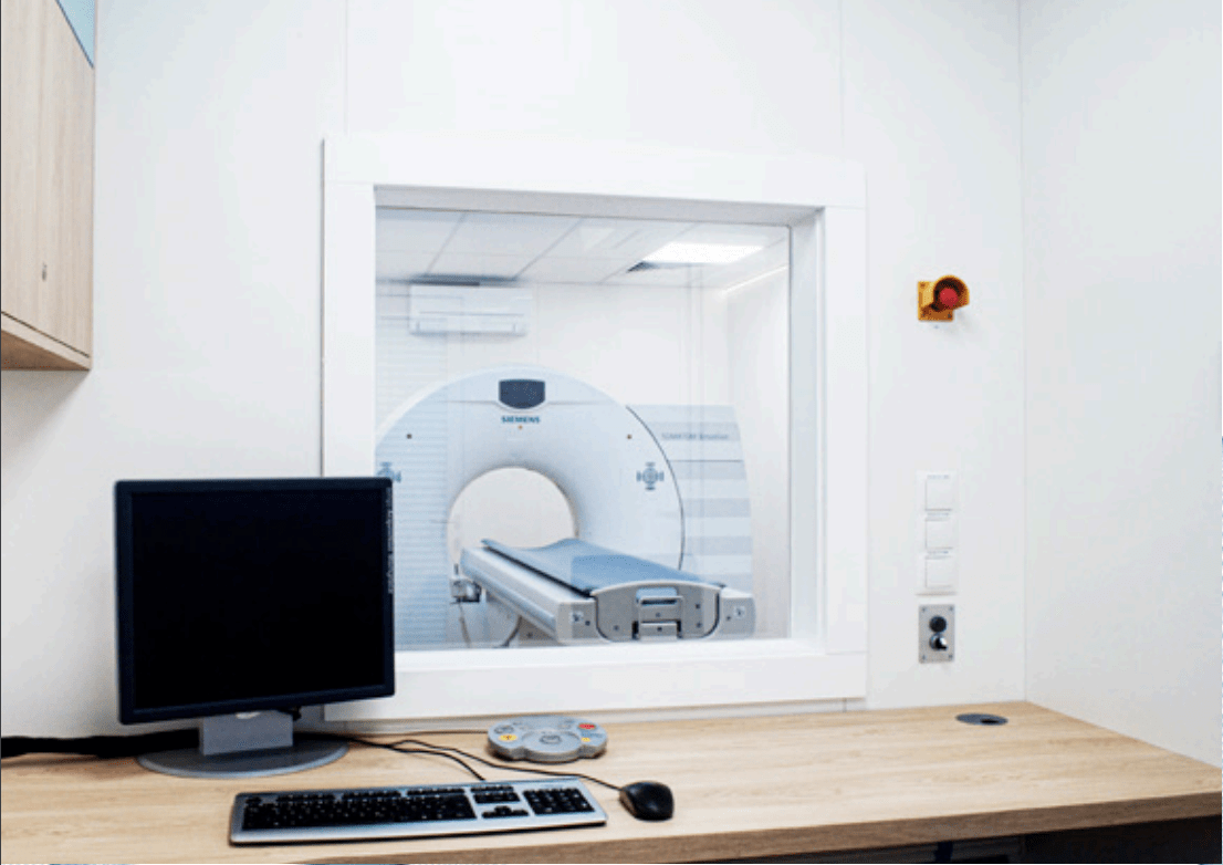
Contrast medium injector
Not included in the rental property and can be purchased separately. Consumables supplied for an additional charge, or provided by the tenant

Consumables
The consumables must be provided by the customer.

OPERATING ROOM

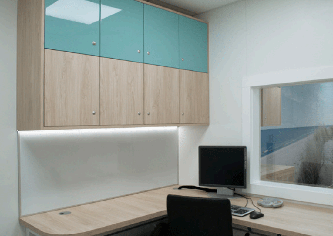
Fitted wardrobes and shelves
for documents, utensils and operating instructions

CT console

Control panel for light and climate
Emergency switch The system has an emergency shutdown system:

De-energizing the system: The system is de-energized in an emergency, which can lead to damage to the system and loss of data.

CT SPECIFICATION

DEVICE: 6-slice CT scanner
MODEL: Siemens® Somatom Emotion 6 #188

CONDITION: excellent – technical & optical
TUBE: Dura 422 MV

Gantry

  • Aperture: 70 cm
  • Scan Field: 50 cm
  • Tilt: +/- 30°
  • Rotational times: 0.6, 0.8, 1.0, 1.5 s

Detector

  • Number of detector rows: 16
  • Elements: 11,776
  • Channels per slice: 1,472
  • Number of projections Up to 1.875 (1/360°)
  • Max. slices / revolution: 6

Image Reconstruction

  • Reconstruction time up to 8 images/s
  • Reconstructed slice widths: 0.63, 0.75, 1.0, 1.25, 2.0, 2.5, 3.0, 4.0, 5.0, 6.0, 8.0, 10.0 mm
  • Slice increment: 0.1-10 mm
  • Pitch factor (Volume Pitch): 0.4-1.8
  • Spiral scan time max.: 100 s
  • Scan length max.: 150 cm

Tube assembly

  • Tube DURA: 422MV
  • Tube current range: 20-240 mA
  • Tube voltages: 80, 110, 130 kV
  • Anode heat storage capacity: 5.0 MHU
  • Focal spot size according to: 0.5 x 0.5 mm/7°
  • IEC 60336: 0.8 x 0.7 mm/7°

Patient Table

  • Max. table load: 200 kg/440 lbs
  • Table speed: 1-100 mm/s
  • Vertical table travel range: 45-83 cm (at table top)
  • Vertical travel speed: <22.4 mm/s
  • Scanable range (metal-free): 153 cm

Installation

  • Examination room Temperature range: 18-30° C
  • Heat dissipation gantry: max. 6.8 kW
  • Surface area for installation: 18 m2
  • Power supply: 380-480 V
  • Nominal line frequency: 50; 60 Hz
  • Nominal power connection: <70 kVA
  • Power consumption System on standby: <3.7 kW
  • Power consumption Scanning: <7.0 kW

Included specifications

2D post processing:

  • Image zoom and pan
  • Image manipulations
    – Averaging, subtraction
    – Reversal of gray-scale values
    – Mirroring
  • Advanced image algorithms
    – Low Contrast Enhancement for improving low contrast detectability
    – High Contrast Enhancement for increased sharpness of high contrast structures
    – Advanced Smoothing Algorithm edge preserving and smoothing filter, dedicated to cardiac exams

CARE Dose4D:

  • Achieve the best diagnostic image quality at lowest possible dose, independent of patient size and anatomy.
  • Fully automated dose management for adults and children with up to 68 % dose reduction.

CARE Filter:

  • Specially designed X-ray exposure filter installed at the tube collimator. Up to 25 % dose reduction with increased image quality.

CINE Display:

Display of image sequences

  • Interactively with mouse controlled rate or automatically
  • Max. image rate >10/s

CT angiography:

  • MIP: Maximum Intensity Projection
  • MinIP function for projection within a small slab to focus on particular vascular structure
  • Evaluation of spiral images and display of vessels, vascular anomalies, aneurysms, plaques, and stenosis

Dynamic Evaluation:

  • Evaluation of contrast enhancement in organs and tissues
  • Calculation of
    – Time-density curves (up to 5 ROI’s)
    – Peak-enhancement images
    – Time-to-peak images

Pediatric protocols:

  • Special clinical protocols with 80 or 110 kV selection and a wide range of mAs settings. The X-ray exposure is adapted to the child’s (and small adults) weight and age, substantially reducing the effective patient dose.

Real-Time MPR:

  • Real-time multiplanar reformatting of secondary views
  • Variable slice thickness (MPR thick, MPR thin) and distance with configurable default values.
  • Viewing perspectives
    – Sagittal
    – Coronal
    – Oblique
    – double oblique
    – freehand (curvilinear)

SureView:

  • Multislice Spiral Image Reconstruction
  • Brilliant image quality and dose savings up to 20 % in spiral mode.

Evaluation Tools:

  • Parallel evaluation of more than10 Regions of Interest (ROI)
    – Circle
    – Irregular
    – Polygonal
  • Statistical evaluation
    – Area/Volume
    – Standard deviation
    – Mean value
    – Min/max values
    – Histogram
  • Profile cuts
    – Horizontal
    – Vertical
    – Oblique
  • Distance measurement
  • Angle measurement
  • Online measurement of a 5 x 5 pixel size ROI
  • Freely selectable positioning of coordinate system
  • Crosshair
  • Image annotation and labeling

Image Transfer / Networking:

Interface for transmitting medical images and information in the DICOM industrial standard. Permits communication between devices from different manufacturers.

  • DICOM Storage (send/receive)
  • DICOM Query/Retrieve
  • DICOM Basic print
  • DICOM Get worklist (HIS/RIS)
  • DICOM MPPS (Modality Performed Procedure Step)
  • DICOM Storage Commitment
  • DICOM Viewer on CD

syngo 3D SSD Surface Shaded Display:

  • Three-dimensional display of surfaces with different density values
    -Soft tissue
    – Bone
    – Contrast
    – enhanced vessels

UFC – Ultra Fast Ceramic Detector:

  • Low patient dose. Up to 30 % dose reduction in relation to conventional CT detectors.
  • More power. More or longer spirals due to low mAs requirements for best possible image quality.
  • More speed. Ultra short afterglow. Specially developed for subsecond and multislice applications requiring a higher projection rate.

Extra specifications:
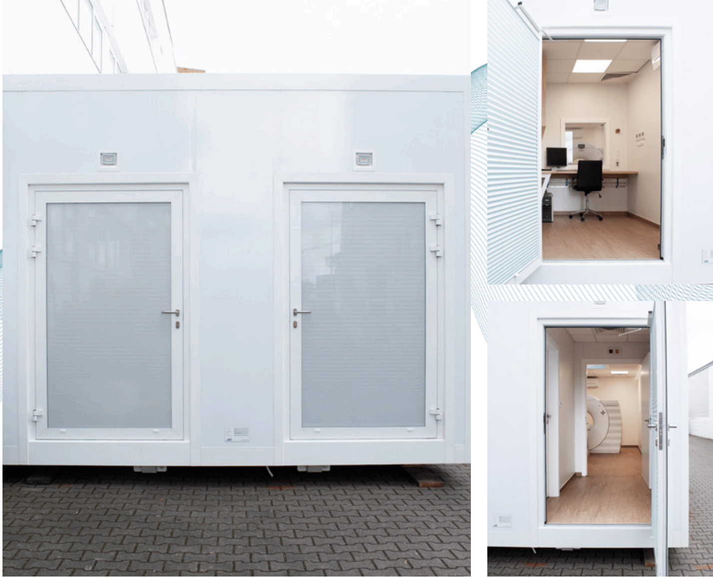
  • Dicom Worklist
  • 3D shaded surface display
  • 3D interactive minimum intensity projection
  • 3D multiplanar reformatting
  • 3D volume editor
  • 3D advanced editor
  • Dental Evaluation
  • Dose modulation
  • Syngo general license
  • Volume rendering
  • HIGH_SPEED
  • Dicom modality performed procedure step
  • RTD
  • HI_RES
  • HIGH_POWER
  • CAP3D_BONEREMOVAL
  • DICOM_SR_VIEWER
  • EMOTION6
  • Biopsy_Enh.
  • CARE Vision

LOCATION REQUIREMENTS

Area to be provided

An on-site inspection is recommended to determine the access route and the area to be provided. The area to be provided is 13.00 m x 5.50 m (l x w). There should also be sufficient space for transporting the bed.

Underground

In principle, a solid base with a load-bearing capacity of 25.0 t is required. Additional reinforcement is required if the substrate is unstable. This can be done, for example, with concrete, paving or road surfacing (bound materials). The forecourt required to allow patient access to the CT scanner via the front entrance door does not need to be specially reinforced.

Installation

With the help of an additional truck crane, the building is craned from the low-loader truck to the appropriate location. The additional space required for the crane must be taken into account here. (The costs for the crane rental shall be borne by the tenant).

Site characteristics

The floor must be level and horizontal in order to be able to position the system easily and ensure trouble-free operation of the system. Unevenness should not exceed a slope of 1%. Please contact MEDSER Medical Services in the event of major unevenness, slopes or inclines.

Access route

The access road to the site must be passable for delivery and removal with a low-loader including tractor unit (kerbs, wall projections, free of snow and ice in winter). Furthermore, the customer must ensure that no parked cars obstruct delivery and collection. If a crane is required on site, sufficient space must be available for the crane truck.

Craning

  • The container must be lifted using 4 12T eyelets, which are attached to the floor frame (Fig. a) ).
  • The craning must be as shown in the pictures b) & c) must take place:
  • The container should not be lifted at wind speeds above 10 m/s.

It is forbidden,

  • stay inside or on the upper levels of the container.
  • to move the container under high-voltage power lines.
  • stay under the boom when lifting and moving the container suspended from the hooks.
  • to move the container over people and the driver’s compartment.
  • to leave the container hanging on the hook unattended after work or during breaks.
  • to lift the container with unsecured equipment inside.

The container weight is approximately: 22.7t

Footprint:
The container must rest on at least 8 points of 90 x 90 cm.

LOCATION REQUIREMENTS

Electrical connections

  • Power supply: 400 V – Frequency: 50 Hz
  • Power: 100 kVA
  • Standard connection plug: 125A CEE plug
  • Power connection cable: Must be provided by the customer
  • Voltage deviation: +/- 5 %
  • Frequency deviation: +/- 0.5 Hz
  • Rotating field of the customer-side connection: clockwise
  • Residual current circuit breaker is integrated in the container main circuit.

Network

  • The system has a network connection for sending image data and for saving or printing images in DICOM 3.0 format.
  • The system-side network socket (type CAT 5) is located in the connection box (at the rear in the direction of travel).
  • The end sockets are located in the control room.
  • The customer must provide a connection via a freely accessible network socket (CAT 5 socket, RJ45 plug) near the location or at the power connection.
  • Any measures (hardware and software related) required to connect the system to the network are the responsibility of the customer.
  • The network cable must be provided by the customer.

Phone

  • The phone must be provided by the user

ANFRAGE

CT-CUBE 1 MOBILE CT-EINHEIT

INQUIRY

CT-CUBE 1 MOBILE CT UNIT