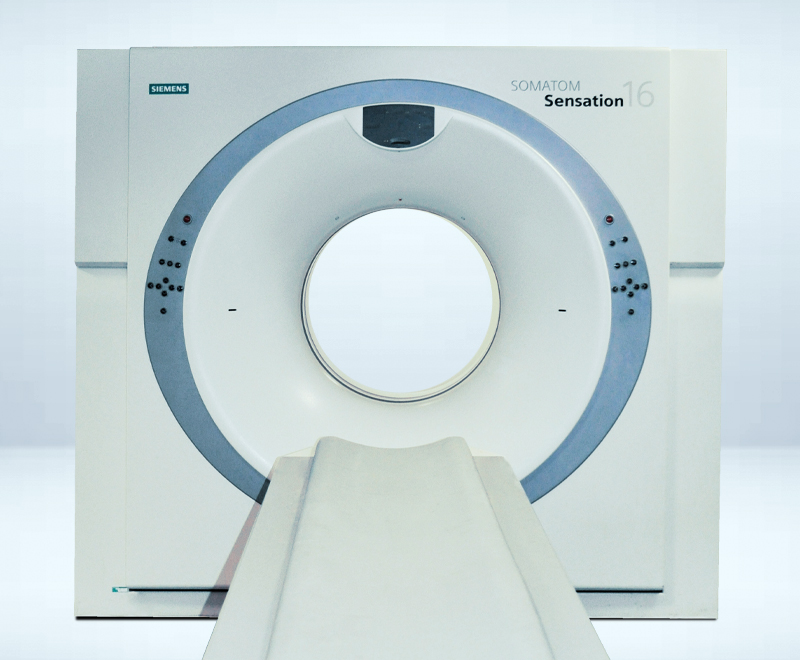
SIEMENS® SOMATOM SENSATION 16 (#587)
(16-ZEILER)

Baujahr: 2005
Zustand: wiederaufbereitet durch MEDSER
Standort: MEDSER Showroom, Heusenstamm/Frankfurt
Röhre: Straton

GANTRY

Aperture78 cm
Scan Field50 cm
Tilt+/- 30°
Rotational times0.42, 0.5, 0.75, 1.0, 1.5 s
Temporal resolutiondown to 92 ms


DETECTOR

Number of detector rows24
Elements16.128
Channels per slice1344
Number of projectionsup to 2.320 (1/360°)
Max. slices / rotation16


IMAGE RECONSTRUCTION

Reconstruction timeUp to 10 images/s
Reconstruction slice widths0.6, 0.75, 1.0, 1.5, 2.0, 3.0, 4.0, 5.0, 6.0, 7.0, 8.0,10.0 mm
Slice increment0.1–10 mm
Pitch factor (Volume Pitch)0.5–2.0 (1–32)
Scan lengthmax. 157 cm
Spiral scan timeMax. 100 s


TUBE ASSEMBLY

TubeSTRATON
Tube current range28–580 mA
Tube voltages80, 100, 120, 140 kV
Focal Spot size0.6 x 0.7 mm/7°


PATIENT TABLE

Max. table load200 kg
Table speed1–150 mm/s
Vertical table travel range53–102 cm (at table top)
Vertical travel speed2.5-45 mm/s
Scanable range (metal-free)157 cm


INSTALLATION

Examination room Temperature range15-28°„ C
Heat dissipation gantrymax. 15 kW
Surface area for installation30 m2
Power supply380-480 V
Nominal line frequency50; 60 Hz
Max. power connection66–83 kVA (fuse 100 A)
Power consumption Computer on2 kW
Power consumption System on standby8 kW
Power consumption Scanning50,0 kW


INCLUDED SPECIFICATIONS

2D post processing

  • Image zoom and pan
  • Image manipulations
    • Averaging, subtraction
    • Reversal of gray-scale values
    • Mirroring
  • Advanced image algorithms
    • Low Contrast Enhancement for improving low contrast detectability
    • High Contrast Enhancement for increased sharpness of high contrast structures
    • Advanced Smoothing Algorithm edge preserving and smoothing filter, dedicated to cardiac exams

CARE Bolus CT

  • Scan mode for contrast bolus triggered data acquisition.
  • Significant improvement of the planning procedure and diagnosis by enabling an optimum spiral scan start after contrast injection.
  • The procedure is based on repetitive low-dose monitoring scans at one slice level and analysis of the time density curve in a region-of-interest (ROI).

CARE Dose

  • Achieve the best diagnostic image quality at lowest possible dose, independent of patient size and anatomy.
  • Fully automated dose management for adults and children with up to 68% dose reduction.

CARE Filter

  • Specially designed X-ray exposure filter installed at the tube collimator. Up to 25% dose reduction with increased image quality.

CARE Topo

  • Real-time topogram
  • Manual interruption possible once desired anatomy has been imaged

CINE Display

  • Display of image sequences
  • Interactively with mouse controlled rate or automatically
  • Max. image rate 30 frames/s

CT-Angiography

  • MIP: Maximum Intensity Projection
  • MinIP function for projection within a small slab to focus on particular vascular structure
  • Evaluation of spiral images and display of vessels, vascular anomalies, aneurysms, plaques, and stenosis

Evaluation Tools

  • Parallel evaluation of more than10 Regions of Interest (ROI)
    • Circle
    • Irregular
    • Polygonal
  • Statistical evaluation
    • Area/Volume
    • Standard deviation
    • Mean value
    • Min/max values
    • Histogram
  • Profile cuts
    • Horizontal
    • Vertical
    • Oblique
  • Distance measurement
  • Angle measurement
  • Online measurement of a 5 x 5 pixel size ROI
  • Freely selectable positioning of coordinate system
  • Crosshair
  • Image annotation and labeling

Image Transfer / Networking

Interface for transmitting medical images and information in the DICOM industrial standard. Permits communication between devices from different manufacturers.

  • DICOM Storage (send/receive)
  • DICOM Query/Retrieve
  • DICOM Basic print
  • DICOM Get worklist (HIS/RIS)
  • DICOM MPPS (Modality Performed Procedure Step)
  • DICOM Storage Commitment
  • DICOM Viewer on CD

Pediatric protocols

  • Special clinical protocols with 80 or 110 kV selection and a wide range of mAs settings. The X-ray exposure is adapted to the child’s (and small adults) weight and age, substantially reducing the effective patient dose.

SureView

  • Multislice Spiral Image Reconstruction
  • Brilliant image quality and dose savings up to 20% in spiral mode.

syngo 3D SSD
(Surface Shaded Display)

  • Three-dimensional display of surfaces with different density values
    • Soft tissue
    • Bone
    • Contrast-enhanced vessels

syngo Dynamic Evaluation

  • Evaluation of contrast enhancement in organs and tissues
  • Calculation of
    • Time-density curves (up to 5 ROI’s)
    • Peak-enhancement images
    • Time-to-peak images

UFC – Ultra Fast Ceramic Detector

  • Low patient dose. Up to 30% dose reduction in relation to conventional CT detectors.
  • More power. More or longer spirals due to low mAs requirements for best possible image quality.
  • More speed. Ultra short afterglow. Specially developed for sub-second and multislice applications requiring a higher projection rate.

EXTRA SPECIFICATIONS 

  • 3D advanced editor
  • 3D Boneremoval
  • 3D interactive minimum intensity projection
  • 3D multiplanar reformatting
  • 3D shaded surface display
  • 3D volume editor
  • 3D Volume Rendering
  • Bolus-Track
  • Cardiac Speed 037
  • Cardiac Speed 04
  • Cardio Turbo Recon
  • CARE Dose Modulation
  • CARE Vision
  • Dicom modality performed procedure step support
  • Dicom Worklist
  • High Speed
  • Osteo Evaluation
  • Perfusion
  • Power Tube
  • Volume Workflow

ANFRAGE

SIEMENS® SOMATOM SENSATION 16 (#587) (16-ZEILER)