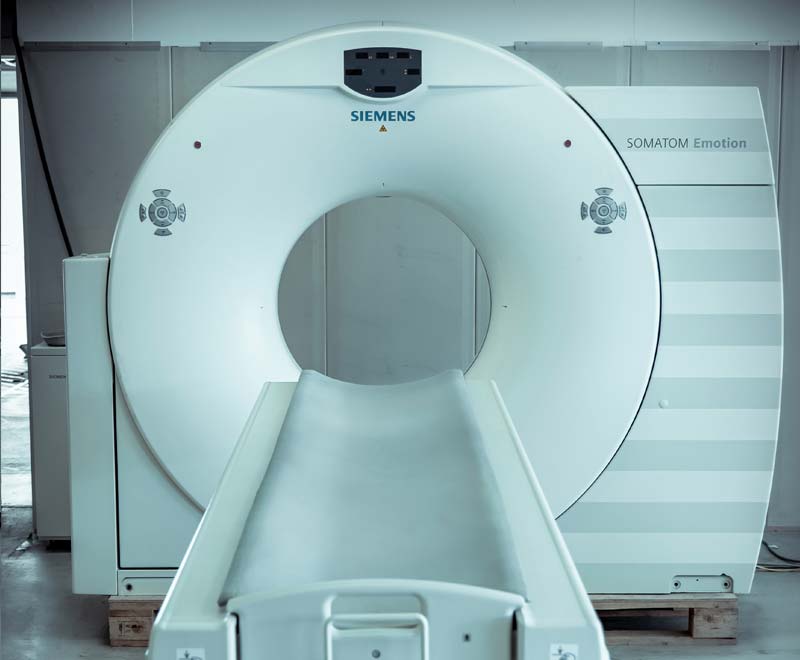
SIEMENS® SOMATOM EMOTION 16 (#907)
(16-SLICE SCANNER)

Manufactured: 2010
Condition: reconditioned by MEDSER
Location: MEDSER Showroom, Heusenstamm/Frankfurt
Tube: Dura 422 MV

GANTRY

Aperture 70 cm
Scan Field 50 cm
Tilt +/- 30°
Rotational times 0.6, 1.0, 1.5 s

DETECTOR

Number of detector rows 16
Elements 17.664
Channels per slice 1472
Number of projections Up to 1,250 (1/360°)
Max. slices / rotation 16

IMAGE RECONSTRUCTION

Reconstruction time up to 8 images/s
Reconstruction slice widths 0.6, 0.75, 1.0, 1.5, 2.0, 3.0, 4.0, 5.0, 6.0, 8.0, 10.0 mm
Slice increment 0.1-10 mm
Pitch factor (Volume Pitch) 0.4-1.8
Spiral scan time max. 100 s
Scan length max. 150 cm

TUBE ASSEMBLY

Tube DURA 422MV
Tube current range 20-345 mA
Tube voltages 80, 110, 130 kV
Anode heat storage capacity 5.0 MHU
Focal Spot size according to IEC 60336 0.8 x 0.5 mm/7°
0.8 x 0.7 mm/7°

PATIENT TABLE

Max. table load 220 kg
Table speed 1-100 mm/s
Vertical table travel range 45-83 cm (at table top)
Vertical travel speed <22.4 mm/s
Scanable range (metal-free) 153 cm

INSTALLATION

Examination room Temperature range 18-30° C
Heat dissipation gantry max. 6.8 kW
Surface area for installation 18 m2
Power supply 380-480 V
Nominal line frequency 50; 60 Hz
Max. power connection <70 kVA
Power consumption System on standby <3.7 kW
Power consumption scanning <7.0 kW

INCLUDED SPECIFICATIONS

2D post processing

  • Image zoom and pan
  • Image manipulations
    • Averaging, subtraction
    • Reversal of gray-scale values
    • Mirroring
  • Advanced image algorithms
    • Low Contrast Enhancement for improving low contrast detectability
    • High Contrast Enhancement for increased sharpness of high contrast structures
    • Advanced Smoothing Algorithm edge preserving and smoothing filter, dedicated to cardiac exams

CARE Bolus CT

  • Scan mode for contrast bolus triggered data acquisition.
  • Significant improvement of the planning procedure and diagnosis by enabling an optimum spiral scan start after contrast injection.
  • The procedure is based on repetitive low-dose monitoring scans at one slice level and analysis of the time density curve in a region-of-interest (ROI).

CARE tin

  • Automated real-time tube current adjustment for best diagnostic image quality at lowest possible dose, independent of patient size and anatomy
  • Fully automated dose management for adults and children with up to 68% dose reduction

CARE Filter

Specially designed X-ray exposure filter installed at the tube collimator. Up to 25% dose reduction with increased image quality.

CARE Topo

  • Real-time topogram
  • Manual interruption possible once desired anatomy has been imaged

CINE Display

  • Display of image sequences
  • Automatic or interactive with mouse control
  • Max. image rate >10 frames/s

CT angiography

  • MIP: Maximum Intensity Projection
  • MinIP function for projection within a small slab to focus on particular vascular structure

    Evaluation of spiral images and display of vessels, vascular anomalies, aneurysms, plaques, and stenosis

Evaluation Tools

  • Parallel evaluation of more than10 Regions of Interest (ROI)
    • Circle
    • Irregular
    • Polygonal
  • Statistical evaluation
    • Area/Volume
    • Standard deviation
    • Mean value
    • Min/max values
    • Histogram
  • Profile cuts
    • Horizontal
    • Vertical
    • Oblique
  • Distance measurement
  • Angle measurement
  • Online measurement of a 5 x 5 pixel size ROI
  • Freely selectable positioning of coordinate system
  • Crosshair
  • Image annotation and labeling

Image Transfer / Networking

  • Interface for transmitting medical images and information in the
    DICOM industrial standard. Permits communication between devices from different manufacturers.
  • DICOM Storage (send/receive)
  • DICOM Query/Retrieve
  • DICOM Basic print
  • DICOM Get worklist (HIS/RIS)
  • DICOM MPPS (Modality Performed Procedure Step)
  • DICOM Storage Commitment
  • DICOM Viewer on CD

Pediatric protocols

  • Special clinical protocols with 80 or 110 kV selection and a wide range of mAs settings. The X-ray exposure is adapted to the child’s (and small adults) weight and age, substantially reducing the effective patient dose.

SureViewTM

  • Multislice Spiral Image Reconstruction
  • Brilliant image quality and dose savings up to 20% in spiral mode.

syngo 3D SSD
(Surface Shaded Display)

  • Three-dimensional display of surfaces with different density values
    • Soft tissue
    • Bone
    • Contrast-enhanced vessels

syngo Dynamic Evaluation

  • Evaluation of contrast enhancement in organs and tissues
  • Calculation of
    • Time-density curves (up to 5 ROI’s)
    • Peak-enhancement images
    • Time-to-peak images

UFC – Ultra Fast Ceramic Detector

  • Low patient dose. Up to 30% dose reduction in relation to conventional CT detectors.
  • More power. More or longer spirals due to low mAs requirements for best possible image quality.
  • More speed. Ultra short afterglow. Specially developed for sub-second and multislice applications requiring a higher projection rate.

EXTRA SPECIFICATIONS

  • Real-Time MPR

    • Real-time multiplanar reformatting of secondary views
    • Variable slice thickness (MPR thick, MPR thin) and distance with configurable default values.
    • Viewing perspectives
      • Sagittal
      • Coronal
      • Oblique
      • double oblique
      • freehand (curvilinear)
  • RTP Enhancement

    • Hardware and software components to optimize the RTP process

syngo CT Oncology

  • Fast-track routine diagnostic oncology, staging, and follow-up. It provides a range of fully automated tools specifically designed to support physicians in the detection, segmentation, and evaluation of suspicious lesions including dedicated tools for lung, liver, and lymph node assessment. It also offers a fully automated follow-up protocol and features LungCAD (Computer Assisted Detection). syngo CT Oncology also facilitates functional imaging offering fusion of PET with CT data.

syngo Dental CT

  • Reformatting of panoramic slices and paraxial sections through the lower and upper jaw for analysis in connection with implantation surgery

syngo InSpace4D

  • One-click bone removal
  • One-click table removal
  • Automated segmentation and removal of bony structures for vascular analysis
  • 4D evaluation of the beating heart with full resolution
  • Real-time navigation through moving anatomy in user selectable arbitrary planes
  • High performance volume reading for physician’s diagnosis and pre-surgical planning in daily clinical routine

syngo LungCARE CTa

  • Software for fast 3D-based visualization and quantitative evaluation of lung nodules, with lowest possible radiation dose. Includes fully automated follow-up.

syngo Osteo CT

  • Non-invasive measurement of the bone mineral density of the lumbar spine to help early diagnosis of osteopenia and osteoporosis, and to assess the effectiveness of treatment
  • Osteo CT measurements are standardized to the ESP Phantom (ESP: European Spine Phantom)
  • Includes table mat and reference phantom for Osteo CT studies

WorkStream4D

  • 4D workflow with direct generation of axial, sagittal, coronal, or double-oblique images from standard scanning protocols
  • Elimination of manual reconstruction steps
  • Reduction of data volume up to a factor of 10, since virtually all diagnostic information is captured in 3D slices

More specifications

  • CAP3D Boneremoval
  • CAP3D Filter SSD
  • CAP3D Filter MIP
  • CAP3D Main
  • CAP3D Voimode
  • CAP3D Track
  • CAP3D Filter VRT
  • CAP3D Editor
  • CARE tin
  • MPPS Support
  • Volume Workflow
  • RTD
  • Bolus Track
  • Syngo General License
  • DICOM SR Viewer
  • MR Support i
  • Get Worklist

ANFRAGE

SIEMENS® SOMATOM EMOTION 16 (#907) (16-ZEILER)

INQUIRY

SIEMENS® SOMATOM EMOTION 16 (#907) (16-DIVISION)